Views: 0 Author: Site Editor Publish Time: 2026-06-06 Origin: Site
Small dental practices face a constant financial squeeze today. You must carefully balance tight capital budgets against the undeniable need to modernize your clinical technology. The central question remains whether a digital imaging tool is merely a "nice-to-have" gadget. Or is an Intraoral Camera a foundational tool designed for practice growth and patient retention? Every equipment dollar you spend must deliver clear, measurable value to your business.
We provide a skeptical, evidence-based breakdown of the real-world returns these devices offer. You will discover the implementation risks and the exact decision criteria necessary for evaluating this hardware. Many practices buy into marketing hype without understanding the operational impact. Prepare to uncover what truly drives profitability, workflow efficiency, and patient trust in modern dentistry.
Direct vs. Indirect ROI: Intraoral cameras rarely generate direct billable revenue; their financial value lies in significantly higher case acceptance rates through patient co-diagnosis.
Operational Efficiency: High-quality clinical imaging reduces dental insurance claim denials by providing irrefutable visual evidence of structural damage (e.g., fractures, marginal breakdown).
Adoption Hurdles: The most common point of failure is lack of staff training and poor integration with existing Practice Management Software (PMS).
Evaluation Focus: Practice owners should prioritize software compatibility, ease of use for hygienists, and durability over raw megapixel counts.
Modern dentistry relies heavily on patient education. Transitioning your practice from a "tell" methodology to a "show" methodology fundamentally shifts the patient relationship. When you simply describe a cracked amalgam filling, patients often hesitate. They may doubt the necessity of an expensive crown if they currently feel no pain. However, displaying a highly magnified, illuminated image of a leaking margin directly on a monitor removes that skepticism.
This visual evidence builds immediate trust. It significantly reduces the psychological friction patients feel when evaluating restorative treatments. We call this process "patient co-diagnosis." The patient effectively sees what you see. They become active participants in their own treatment planning. The indirect return on investment becomes clear when your acceptance rates for large cases climb. An investment in high-quality Intraoral Cameras easily pays for itself after just a few accepted crown or bridge cases.
Dental insurance clearinghouses routinely reject claims lacking sufficient clinical narratives. Relying solely on written descriptions and standard radiographs often results in delayed payments. Clear intraoral photography provides undeniable visual proof of structural damage. If a tooth exhibits a severe fracture line that an X-ray fails to capture, a high-resolution color photo fills the evidence gap.
Attaching these images to your claims directly reduces rejection rates. You eliminate the back-and-forth appeals process with insurance adjusters. This accelerated approval cycle drastically improves your accounts receivable workflow. Faster claim processing means steadier cash flow for a small practice.
Many new practice owners wonder how to bill for these digital images. The straightforward reality is you typically do not charge for intraoral photos. You must view the camera as a diagnostic and communication tool, not a separate line-item procedure. Attempting to bill patients for routine photos creates unnecessary friction. It undermines the trust you intend to build. The true financial value exists in the treatments the photos help you sell, alongside the administrative time you save on insurance appeals.
Traditional bite-wing X-rays remain essential, yet they possess distinct limitations. They easily miss early-stage pathologies. High-intensity LED illumination and macro-magnification change the diagnostic landscape. You can instantly detect micro-fractures, secondary caries hiding beneath old composites, and subtle leaking margins.
A magnified image reveals structural compromises invisible to the naked eye. This clarity allows you to intercept dental issues before they escalate into painful emergencies. The precision protects your patients and elevates your standard of clinical care.
Your hygienists represent the frontline of patient communication. Integrating this imaging tool into routine recall appointments yields massive benefits. A hygienist can quickly capture a "tour of the mouth" during a standard prophylaxis visit. This creates a highly accurate visual baseline.
Over time, you can monitor wear patterns, tissue changes, and restorative degradation. Comparing a photo from today with one from two years ago is powerful. Patients immediately recognize the progression of recession or enamel wear. The hygienist warms up the patient to necessary treatments before the dentist even enters the operatory for the exam.
Thorough visual documentation serves as a critical medico-legal shield. Comprehensive charting protects small practices from liability claims. When patient files contain detailed, date-stamped photographs, you demonstrate a rigorous adherence to clinical standards. This level of documentation heavily supports E-E-A-T principles—Experience, Expertise, Authoritativeness, and Trustworthiness—within your clinical protocols.
Best Practice: Always attach a brief, standardized note to every saved image in your software. A photo labeled "Upper Right Quadrant - Tooth 3 distolingual fracture" carries much more weight than an unnamed image file.
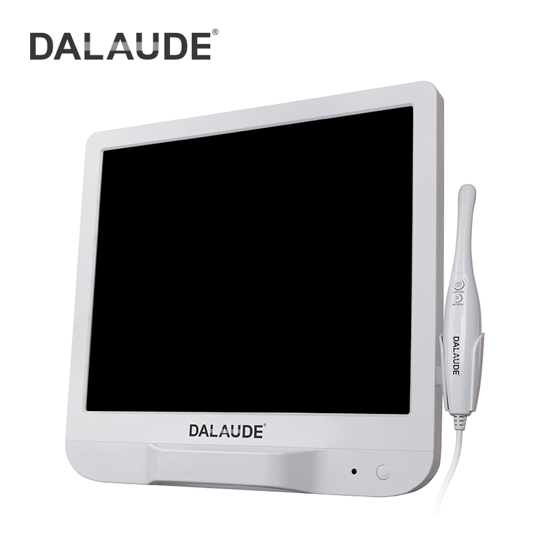
Many practice owners severely underestimate software compatibility. Purchasing a device solely based on its physical specs is a major mistake. Proprietary software environments often trap clinics into frustrating workflows. If your new camera forces you to open a separate imaging program outside of your Practice Management Software (PMS), your staff will hate using it.
You must verify strict TWAIN compatibility. TWAIN acts as a universal communication protocol between imaging hardware and software platforms. Seamless integration with systems like Dexis, Eaglesoft, or Open Dental is non-negotiable. The image should capture and save directly into the patient's active chart with a single button press. Anything less creates an administrative bottleneck.
Operational annoyances frequently derail technology adoption. If a tool frustrates a dental assistant, it ends up sitting in a drawer. Watch out for these common friction points:
Fogging Lenses: Rapid temperature changes in the mouth cause cheap lenses to fog instantly. Staff lose valuable minutes waiting for defogging.
Difficult Focal Adjustments: Cameras requiring manual focus wheels are clumsy to operate while holding a mirror and retracting a cheek.
Cumbersome USB Cords: Thick, rigid cables pull on the operator's wrist. Frequent bending causes internal wires to fray and fail prematurely.
Button Placement: Capture buttons placed awkwardly lead to blurry photos because the user inevitably shakes the wand when pressing them.
Hardware sits unused if hygienists and assistants find it disruptive. Dental appointments run on strict timelines. A prophy visit leaves little margin for error. If capturing images adds five minutes to an appointment, the staff will simply skip the step.
Proper training is a hidden, mandatory cost. You cannot merely drop a new camera in the operatory and expect immediate adoption. Dedicate specific time for hands-on practice. Let your team practice capturing images on each other until the workflow feels like second nature. When your team feels confident, patient implementation becomes frictionless.
Choosing between wired and wireless models impacts your daily operations significantly.
Wired Models: These are generally more reliable. They offer a constant power supply and steady data transmission. They typically cost less and weigh less. The downside is cord management and the eventual degradation of the physical connection.
Wireless Models: These units offer fantastic ergonomic freedom. You can easily pass them between operatories. However, they introduce battery management realities. Forgetting to charge the unit paralyzes the workflow. They also carry a heavier physical footprint due to internal batteries.
The lens capability dictates your image quality and ease of use. Fixed-focus lenses boast a lower learning curve. They stay in focus within a specific distance range. They are incredibly easy for new staff to pick up and use immediately.
Conversely, variable or liquid-lens autofocus models deliver superior image quality. They adapt to macro shots of a single tooth or wide-arch views seamlessly. However, they require slightly higher user intervention. The wand must momentarily stabilize to lock focus before capture. Consider your staff's technical comfort level when choosing.
Navigating the equipment market requires understanding what you actually pay for. Price directly correlates with software native integration and physical durability.
Market Tier | Price Range | Durability & Integration | Ideal For |
|---|---|---|---|
Budget / Direct-to-Consumer | $100 - $500 | High failure rates. Often requires unstable third-party software patches. Poor lens seals. | Highly risky for long-term clinical use. Not recommended for busy practices. |
Mid-Tier Value | $500 - $1,500 | Solid TWAIN bridging. Acceptable optics and decent physical build quality. | Often the sweet spot for small practices looking to outfit multiple hygiene rooms. |
Premium / Brand-Name | $2,000+ | Highest durability. Native PMS integration without TWAIN glitches. Premium optics. | Practices wanting zero workflow friction, though it carries a longer break-even period. |
We recommend immediate adoption if your practice currently suffers from low restorative case acceptance. If you constantly hear "let me think about it" after presenting treatment plans, you need visual aids. Additionally, if your front desk battles high insurance denial rates for crowns and core buildups, immediate investment is justified. The tool will rapidly resolve both operational pain points.
Technology adoption requires workflow stability. Suggest pausing your purchase if your practice is currently overhauling its core practice management software. Installing new hardware during a major software migration invites chaos. Furthermore, if you are experiencing high staff turnover, delay the purchase. Wait until you secure a stable clinical team. You want to train a permanent team once, rather than constantly retraining a revolving door of assistants.
Do not buy blindly from a catalog. Advise your clinical director or lead assistant to request a multi-day in-office trial from trusted vendors. Test the exact PMS integration on your actual computers. Let your hygienists test the wand's ergonomics during live appointments. Only commit to a purchase when the team confirms the device genuinely improves their clinical flow.
For a small practice, adopting this digital imaging tool is one of the lowest-barrier entry points into modern digital dentistry.
The financial break-even point is achieved rapidly through improved case acceptance, not direct billing.
Never underestimate the importance of TWAIN compatibility; seamless software bridging is more crucial than physical megapixel counts.
Successful implementation relies heavily on dedicated staff training and ergonomic comfort.
Next Step: Audit your recent insurance claim rejections and stalled treatment plans this week. If a visual image could have saved those cases, contact a vendor for a hardware trial immediately.
A: Generally, no. Intraoral photographs (CDT code D0350) are typically only billable if they are explicitly requested by the insurance company to support a specific claim, not for routine screening. You should view the images as diagnostic aids that help secure approvals for other major treatments.
A: Depending on the tier and usage, commercial-grade cameras typically last 3 to 5 years. Cord degradation and sensor failure are the most common end-of-life causes. Proper handling and careful cord storage greatly extend the device's lifespan in a busy practice.
A: Yes. Patient education literature heavily supports that seeing their own oral condition in real-time drastically improves trust. It validates the dentist's recommendations, removes the mystery of dental terminology, and significantly reduces the perceived pressure of case presentation.