Views: 0 Author: Site Editor Publish Time: 2026-06-05 Origin: Site
The primary bottleneck in preventive dentistry rarely involves a clinician’s diagnostic skill. Instead, the gap typically forms between an accurate clinical diagnosis and the patient ultimately accepting the proposed treatment plan. Dentists can easily spot early signs of decay, but communicating this urgency to a layman remains surprisingly difficult.
Modern dental practices are currently shifting away from verbal explanations. They now rely heavily on objective visual evidence. Patients grow increasingly skeptical of recommended procedures they cannot physically see in their own mouths. They often dismiss early interventions as unnecessary upselling or aggressively question the need for proactive care.
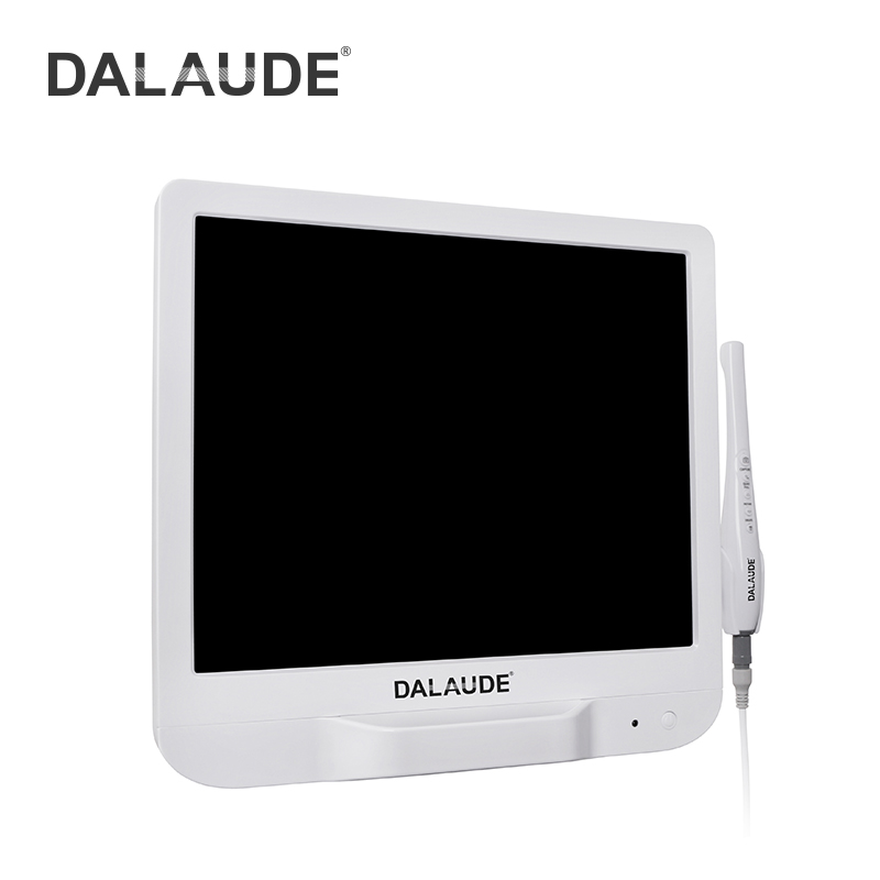
Integrating a modern Intraoral Camera transitions your dental practice from simply "telling" a patient about an issue to clearly "showing" them. You will discover how verifiable visual proof directly impacts preventive care compliance, elevates clinical documentation accuracy, and ultimately strengthens overall practice revenue.
Visual Evidence Drives Acceptance: High-resolution imagery turns abstract dental issues (e.g., micro-fractures, early decay) into undeniable visual proof, increasing preventive treatment acceptance rates.
Integration is Critical: The most capable hardware fails without seamless integration into existing Practice Management Systems (PMS) and imaging software.
Workflow Adoption > Features: Successful implementation relies on ergonomic hardware and low-friction staff training, not just maximum megapixel counts.
Security & Compliance: Modern cloud-connected cameras require evaluation through a strict HIPAA and data security lens.
Dental practices lose thousands of dollars annually due to unaccepted treatment plans. When patients decline preventive interventions, dentists often adopt a "watch and wait" approach. This passive strategy creates delayed care scenarios. It also harms practice revenue. Patients ignore developing micro-fractures or early carious lesions until pain forces them into the chair. This cycle damages patient trust and turns routine checkups into high-stress emergency visits.
A successful equipment deployment completely changes this dynamic. Clinical success requires clear benchmarks. Your team should capture standardized baseline charting for every new patient. You must document lesion progression objectively over multiple visits. Accurate visual records prevent subjective guessing during six-month recalls. Improved communication naturally follows when patients view a monitor displaying their own oral health status.
Visual data disarms inherent patient skepticism. Dentists frequently fight a frustrating trust deficit. Patients often view verbal recommendations as sales pitches. High-resolution imagery bridges this gap instantly. A patient looking at a magnified, cracked amalgam filling on a screen cannot argue against the physical reality. This verifiable evidence moves the interaction away from a transactional negotiation. It creates a collaborative healthcare evaluation where you and the patient tackle the visible problem together.
Vendors constantly push raw megapixel counts in their marketing materials. However, clinical utility relies on depth of field, autofocus reliability, and LED illumination quality. A 4K sensor means very little if the lens cannot maintain focus on a moving patient. Superior depth of field ensures the entire posterior tooth remains sharp. Bright, even LED illumination prevents shadows from obscuring critical details.
You must prioritize macro-focus capabilities above all else. Preventive dentistry depends on capturing early-stage indicators. Clinicians need extreme close-ups of hairline fractures, demineralization spots, and marginal leakage. Look for optical systems specifically designed for close-range diagnostic clarity.
Chart: Optical Features vs. Marketing Myths | ||
Hardware Feature | Clinical Importance | Common Marketing Myth |
|---|---|---|
Depth of Field | Keeps multiple teeth in sharp focus without constant readjustment. | "Higher megapixels automatically mean sharper images." |
Macro-Focus | Reveals early demineralization and micro-fractures clearly. | "Digital zoom works just as well as true optical macro." |
LED Arrays | Eliminates shadows and renders true tissue color. | "Software can perfectly color-correct poorly lit images." |
Choosing between wired and wireless models profoundly impacts your daily operatory flow.
Wired (USB) Architectures: These units provide absolute reliability. You never face battery degradation or unexpected power loss mid-exam. They offer continuous power and zero video latency. However, cable management remains a frustrating issue. Cords often tangle around chairs or limit the hygienist's range of motion.
Wireless Architectures: Wireless models offer unmatched flexibility. You can easily move one unit across multiple operatories. They improve patient comfort by eliminating cords near the face. You must balance this freedom against inherent risks. Battery lifecycles eventually degrade. Video latency can occasionally interrupt smooth scanning. Furthermore, managing charging stations requires strict staff discipline.
Excellent hardware becomes useless without smooth software integration. You must evaluate direct TWAIN compatibility before purchasing any device. Native integrations with dominant platforms like Dentrix, Eaglesoft, or Open Dental save countless hours. Native drivers allow the camera buttons to capture and save images directly into the patient's chart seamlessly.
Common Mistake: Avoid proprietary closed-ecosystem Intraoral Cameras. Some vendors force practices to use their distinct imaging software. This forces your staff to maintain redundant patient databases. It doubles the administrative workload and severely disrupts daily clinical efficiency.
Adding new technology often slows down clinical routines initially. Practice owners must recognize the risk of extending hygiene appointments. The camera should not add ten minutes to a standard checkup. You must outline Standard Operating Procedures (SOPs) to seamlessly integrate image capture into the existing prophy workflow.
Recommended Hygiene SOP:
Capture a full-mouth baseline series during the initial probing phase.
Take targeted macro-shots of any suspicious areas immediately upon discovery.
Leave the images displayed on the overhead monitor while completing the cleaning.
Review the specific images with the patient during the final wrap-up before the dentist enters.
Physical realities dictate daily adoption rates. Ergonomics matter immensely. Lenses fog up when placed against warm mucosal tissue. Heavy wands cause wrist fatigue over an eight-hour shift. Button placement dictates whether a hygienist can operate the device single-handedly while managing a mirror. Infection control also creates friction. You must verify barrier sleeve compatibility. Poorly fitted plastic sheaths degrade image quality drastically, rendering the optical sensor useless.
Staff adoption ultimately determines your success. You must frame training requirements clearly. Hygienists and dental assistants handle the equipment most frequently. They must feel entirely comfortable operating the camera. If the staff finds the device cumbersome, it will sit unused in a drawer.
Modern dental clinics capture massive amounts of sensitive patient data daily. You must evaluate image storage protocols through a strict security lens. Wireless transmission poses unique challenges. Sending high-resolution patient imagery across a local network requires robust encryption. Cloud storage solutions must adhere strictly to HIPAA guidelines. Patient photos, especially those showing identifiable facial features, constitute Protected Health Information (PHI).
Hardware lifecycle management requires realistic planning. Clinical environments are physically demanding. Wands hit the floor. Evaluate drop-resistance ratings carefully before buying. Review the warranty terms regarding accidental damage. You should also verify the availability of replacement parts. Finding a reliable vendor who stocks replacement lenses or USB cables locally prevents extended equipment downtime.
Consistent calibration keeps your diagnostic standards high. Establish a clear maintenance schedule. Sensors and LED arrays degrade slowly over time. This degradation alters color accuracy subtly. A minor color shift might misrepresent healthy gingiva as inflamed tissue. Staff should clean the optical window daily and run software calibration routines monthly to maintain crisp focus mechanisms.
Selecting the right equipment requires a systematic approach. Follow these logical steps to narrow down your options quickly and effectively.
Step 1: Audit Current Infrastructure. Do not look at hardware brochures until you map your current ecosystem. Determine your existing PMS software version. Check your operatory computer specifications. Ensure your graphics cards and USB port types can handle real-time video streaming.
Step 2: Assess Operatory Flow. Decide on your deployment strategy. You can outfit every single room with a budget-friendly wired model. Alternatively, you might purchase one premium wireless unit and rotate it between hygiene rooms. Analyze your daily patient volume to make this decision.
Step 3: Request Clinical Demos. Demand in-office trials from vendors. Testing equipment at a trade show booth is useless. You must test the camera in a real clinical environment. Specifically, check the software integration speeds on your own network. Test how severely barrier sleeves degrade the final image quality.
An intraoral camera acts as a profound investment in patient trust and diagnostic transparency. It serves as far more than just a flashy clinical gadget. It transforms abstract dental terminology into undeniable visual facts. The best devices successfully balance pristine optical clarity with completely frictionless software integration.
Next Steps:
Schedule an immediate IT infrastructure audit to confirm your practice management software compatibility.
Request a live, in-office clinical demonstration from a verified dental technology vendor.
Have your lead hygienist test the ergonomics and barrier sleeve compatibility of at least two competing models.
A: Yes. Visual proof effectively reduces patient friction for out-of-pocket preventive care. When patients physically see a fractured tooth or an open margin, they understand the urgency. This verifiable psychological trigger moves them past skepticism, leading to significantly higher acceptance rates for recommended treatments.
A: Not necessarily. You must distinguish between native integration and basic TWAIN drivers. Native integrations work flawlessly within your software. Standard TWAIN drivers usually work but may require clunky bridging software. Always verify exact compatibility with your specific software version before purchasing.
A: A high-quality wired camera typically lasts three to five years under heavy daily usage. Wireless models may require battery replacements every 18 to 24 months due to charging degradation. Routine maintenance and strict handling protocols significantly extend the overall hardware lifespan.
A: Standard plastic barrier sleeves inevitably introduce some optical distortion and light glare. To mitigate this reality, practices must use custom-fitted sheaths recommended by the manufacturer. High-end cameras are often specifically calibrated to shoot directly through these proprietary sleeves without losing diagnostic clarity.